Tech Project
Description of the challenges faced by the Tech Project
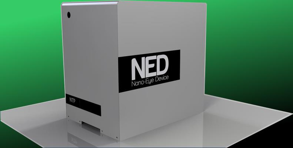
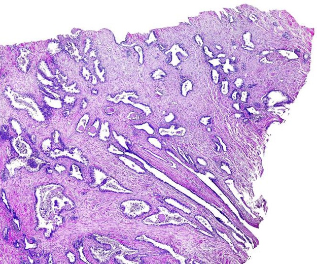
The core challenge of this project is to investigate, develop and propose a solution to better address the pathologist’s transition from the current technologies to the digital ones focusing on the field of Frozen Section Procedure. From a remote location, and in real-time, the pathologist will support the surgical team in the analysis of removed tissues. During the procedure, the slide containing the histological sample will be placed under the smart microscope allowing the specialist to remotely control the microscope, run the clinical images and provide the surgical team with a prompt diagnosis. The tech company will provide its smart microscope platform (NED) and the simulation environment for supporting and implementing the developing the solution for the core challenge.
Brief description of technology
The Digital Pathology is an image-based information environment enabled by computer technology for the management of information generated from a digital slide. Virtual microscopy has permitted the conversion of glass slides into digital ones that can be viewed, managed, shared and analysed on a computer monitor making it, today, one of the most promising avenues of diagnostic medicine to achieve a diagnosis. The Company NTP Nano Tech Projects uses its proprietary smart microscope open platform named NED (Nano-Eye Device), which supports specific pathologist’s activities such as the Intraoperative Examination (known as Frozen Section Procedure). Said platform includes the hardware and software for enabling medical operations where images of biological samples removed from the surgeon (i.e. cancerous tissues) can be captured and analysed in real time by remotely located specialists (pathologists) using different end-user monitors (PC, tablets, smartphones, etc.) without the need of their actual physical presence and ensuring a prompt clinical feedback toward the surgical team before going on with the next surgical steps. This translates into a higher quality of healthcare, an improved efficiency for pathologists and waiting lists, and lesser costs and risks for hospitals in handling glass slides and other resources.
What the project is looking to gain from the collaboration and what kind of artist would be suitable
The objective of the project is to identify a method (it could include a facilitating device or a specific stimulating exercise or an interactive game) that easily and naturally prepares and allows the pathologist the use of remote digital techniques, since the pathologist no longer has to do with the classic analogic microscope with binoculars, but he is far away from the new microscope, the histological sample and the supported surgery team. The artist must know such techniques, realize how the doctor is used to working with the microscope until today, understand the challenge this transition brings about and be able to work alongside the communication networks in order to propose a vision and an implementation that will aid in the project’s fast transition.
Resources available to the artist
The company’s tech team involved in the development of the project will provide the artist with technical support and equipment, the NED smart microscope and the simulation of the proposed clinical activity, Internet connection and access to the laboratory.


