
Tech Project

Description of the challenges faced by the Tech Project
1) Micrometric size-Magnetic Sensors: Arrays of standard MR sensors are fabricated and patterned on silicon substrates. Other substrates can be envisioned including flexible substrates to improve the scanner sensing capabilities while facing topographic irregularities of biological tissues. 2) Thin film deposition: The patterning of materials at nanoscale can be used to design new formats for the MR sensors arrays. The artist will have access to ultra-high vacuum machines. 3) Digital imaging and electronics enable the magnetic/optical signal acquisition for 3D mapping of cells at the molecular scale.
Brief description of technology
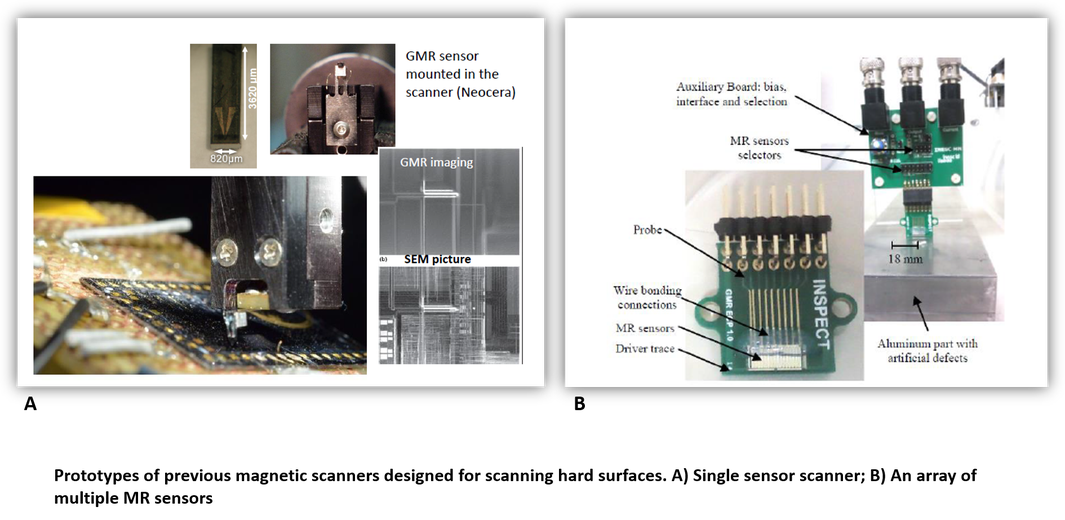
The ability to generate and manipulate the electron spins in magnetic multilayer thin films (with thickness as low as few Å), has led to the development of extremely sensitive magnetic sensors with the capacity to detect ultra-low intensity signals such as those produced by a biomolecule tagged with a single magnetic label. In particular, magnetoresistive (MR) sensors with their tunable response and adjustable operation range are perfect candidates for room temperature applications. Their compatibility with silicon technology and microfabrication enables the production of small foot-print devices, with low power consumption requirements. Moreover, they can be produced at large scale using wafer level processing techniques thus generating low-cost sensing units amenable to be integrated into diagnostic devices. The technology behind MagScopy for Health takes advantage of the consolidated experience of INESC MN on the fabrication of MR sensors for biomedical applications and aims to develop a magnetic camera for a 3D imaging of biological tissues specifically labelled with magnetic nanoparticles for the identification of disease states.
What the project is looking to gain from the collaboration and what kind of artist would be suitable
The project will benefit from a creative point of view in the design of adequate and functional magnetic scanners that can be used by a common pathologist, in standard clinical settings. Features may include for example portability, user-friendly design, proper weight and dimensions. In this view, an artist with and industrial design background and knowledge on material proprieties will be suitable for such propose. The multidisciplinary nature of the project also accommodates other specialties including digital imaging and photography that may address the creation of 3D objects and audio-visual materials (teasers, 3D animations of nanotechnology processes), as follow-up tools of the scientific progress of the project. Alternatively, the possibility to explore new materials at the nano-scale or the optical/magnetic proprieties of biological tissues at the molecular level may convey fine art dedicated artists to explore new perspectives, not usually accessible to the general community.
Resources available to the artist
The INESC-MN facilities will be available to the artists during standard working hours. These will include access to INESC-MN laboratories and Cleanroom once safety measures and training are provided. Whenever equipment usage is necessary an INESC-MN staff member will be always present, including vacuum technologies. The artist will have a dedicated work space including internet access. Additionally, the artist will have access to a variety of materials including metals (aluminum, chromium, gold, titanium, nickel, copper), metal alloys and magnetic materials that can be manipulated at different scales (nm-mm range). Travel budget can be provided.